How to improve microendoscopes? New probe design brings promises to improve biomedical imaging – new paper in IEEE Photonics Journal by Karol Karnowski, PhD
Reading time: about 7 minuts
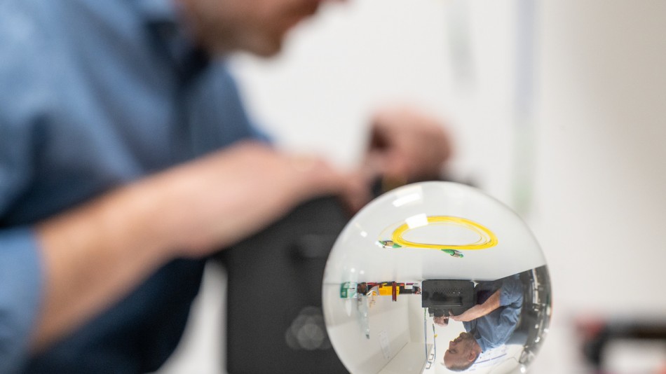
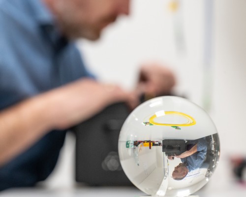
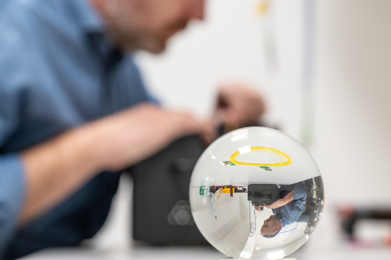
Microendoscopes are the cornerstone of modern medical diagnostics - they allow us to see what we could not even describe two decades ago. The technology is constantly improving, with ICTER scientists contributing to the development of the probes.
Microendoscopes using fiber optics are becoming increasingly important imaging tools, but they have physical limitations. They are essential for applications that require a long working distance, high resolution, and a minimum probe diameter. The research paper titled "Superior imaging performance of all-fiber, two- focusing-element microendoscopes," by Dr. Karol Karnowski of ICTER, Dr. Gavrielle Untracht of the Technical University of Denmark (DTU), Dr. Michael Hackmann of the University of Western Australia (UWA), Onur Cetinkaya of ICTER and Prof. David Sampson of the University of Surrey, sheds new light on modern microendoscopes. It is noteworthy that the research work started while the authors worked in the same research group at UWA.
In it, the researchers showed that endoscopic imaging probes, particularly those for so-called side viewing, combining fiber-optic (GRIN) and spherical lenses, offer excellent performance over the entire range of numerical apertures and open the way to a broader range of imaging applications. In the publication, the performance of endoscopic imaging probes is comparable to commonly used single focusing element probes.
What are microendoscopes?
Miniature fiber-optic probes, or micro-endoscopes, allow imaging of tissue microstructures deep into the specimen or patient. Endoscopic Optical Coherence Tomography (OCT) is particularly promising. It is suitable for volumetric imaging of external tissues and the interior of organs (e.g., the upper respiratory tract, gastrointestinal tract, or lung tubules).
Three main ranges of fiber optic probes can be distinguished. Studies of large, hollow organs (such as those above the upper respiratory tract) require the largest imaging depth ranges (up to 15 mm or more from the probe surface), which can usually be achieved with low-resolution Gaussian beams (spot size in focus in the range of 30-100 μm). The intermediate resolution range (10-30 μm) is helpful for broader applications, such as imaging the esophagus, smaller airways, blood vessels, bladder, ovaries, or ear canal. The biggest challenge is obtaining beams with a resolution better than 10 μm, potentially helpful for animal model studies.
When developing a probe, one must be mindful of design parameters' trade-offs and their impact on imaging performance. Optical systems with a large numerical aperture (high resolution) tend to have a shorter working distance (WD). In addition, better resolution and longer working distance are more difficult to achieve as the probe diameter is reduced. This can be particularly problematic for side viewing probes - a greater minimum working distance is required compared to their forward imaging counterparts. Suppose the probe is encased in a catheter or needle. In that case, this increases the required minimum working distance - in many cases, this is the limiting factor in minimum achievable resolution or probe diameter.
It's worth noting that engineers are usually interested in minimizing the probe diameter for reduced perturbation to the sample and patient comfort. A smaller probe means a more flexible catheter and, therefore, better tolerance of the test by the patient. Thus, one of the best solutions is using monolithic fiber optic probes, whose diameter is limited by the thickness of the optical fibers. Such probes are characterized by ease of fabrication, thanks to fiber-optic welding technology, which avoids the need for tedious alignment and bonding (usually gluing) of individual micro-optical components.
Different types of microendoscopes
The most popular designs of fiber-optic imaging probes are those based on two types of focusing elements: GRIN fiber probes (GFP - GRIN fiber probes) and ball lens probes (BLP - ball lens probes). GRIN probes are easy to make, and their GRIN refractive power is not lost when the refractive index of the surrounding medium is close to that of the fiber used. Commercially available GRIN fibers limit achievable designs. High resolution is tough to achieve with GRIN fibers with small core diameters.
For lateral viewing probes, the curved surface of the fiber (and potentially the catheter) introduces distortion that can adversely affect imaging quality. Spherical BLP probes will not have this problem, but a sphere bigger than the fiber diameter is often required to achieve a resolution comparable to GFP probes. The focusing power of a BLP probe depends on the refractive index of the surrounding medium, which is an important issue when working in a medium with close or near biological samples.
One solution to improve the performance of probes is to use multiple light focusing elements, similar to the design of lenses with a long working distance. Studies have shown that combining numerous light-focusing elements provides better results for many imaging purposes. Probes with multiple focusing elements can achieve better resolution with smaller diameters while offering longer working distances without sacrificing resolution.
How do we improve the probes?
In their latest work, researchers led by Dr. Karnowski have shown that probes with two focusing elements using both GRIN segments and spherical lenses - called GRIN-ball-lens probes (GBLP) - significantly improve the performance of monolithic fiber optic probes. Their first modeling results have already been shown at conferences in 2018 and 2019. GBP probes were compared to the most commonly used GFP and BLP probes and showed performance benefits, especially for applications requiring longer operating distances, better resolution, and small size.
For intuitive visualization of probe performance, the researchers introduced a novel way to comprehensively present simulation results, especially useful when more than two variables are used. Analysis of the effect of GRIN fiber length and spherical lens size led to two interesting conclusions: for optimal results, the range of GRIN fiber length can be kept in the field of 0.25-0.4 pitch length (so-called pitch length); even if the working distance (WD) gain is not so significant for GBLP probes with high numerical aperture, the authors showed that the same or better performance in terms of working distance is achieved for a search with twice the diameter. Moreover, the novel GBLP probes offer higher resolution compared to BLP probes.
The paper's conclusion reads:
We have demonstrated the potential of GBLP probe design for applications with increased working distance, significant for lateral imaging probes, with a highly reduced impact of the refractive index of the probe's environment and a significantly smaller size compared to BLP or GFP probes. These advantages make GBLP probes a tool worth considering for many imaging applications in biological and biomedical research, particularly for projects requiring micro endoscopes.
Author of the press release: Marcin Powęska
Note: The first results from "GRIN-ball-lens probes (GBLP)" modeling have already been shown at the 2018 and 2019 conferences:
- Karol Karnowski, Gavrielle R. Untracht, Michael J. Hackmann, Mingze Yang, Onur Cetinkaya, David D. Sampson, "Versatile, all-fiber, side viewing imaging probe for applications in catheter-based optical coherence tomography," Photonics West, San Francisco, USA, Feb 2019, oral presentation;
- K. Karnowski, G. Untracht, M. Hackmann, M. Yang, O. Cetinkaya, and D. D. Sampson, "Versatile, monolithic imaging probes for catheter-based OCT," 15th Conference on Optics Within Life Sciences, Rottnest Island, Australia, Nov. 2018, poster presentation.
The team responsible for these results started at the University of Western Australia (UWA), and work has now been completed within the following institutions: the Institute of Physical Chemistry, Polish Academy of Sciences and the University of Surrey, one of the authors only remaining at UWA.
Cited paper: K. Karnowski, G. Untracht, M. Hackmann, O. Cetinkaya and D. Sampson, "Superior Imaging Performance of All-Fiber, Two-Focusing-Element Microendoscopes," in IEEE Photonics Journal, vol. 14, no. 5, pp. 1-10, Oct. 2022, Art no. 7152210, doi: 10.1109/JPHOT.2022.3203219.
The International Centre for Translational Eye Research (MAB/2019/12) project is carried out by the Institute of Physical Chemistry, Polish Academy of Sciences within the International Research Agendas programme of the Foundation for Polish Science co-financed by the European Union under the European Regional Development Fund.
Funding sources:
Polish National Agency for Academic Exchange (NAWA) through the Polish Returns Program
University of Western Australia IPRS
Rank Prize Covid Fund
Australian Research Council
University of Surrey


- Author: Marcin Poweska
- Photo source: Karol Karnowski, PhD and Bartłomiej A. Bałamut, MSc
- Date: 27.10.2022


