Magnesium guards vision. An ICTER model identifies a window for retinal protection
Reading time: about 8 minuts
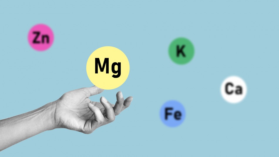
The cells that transmit visual information from the eye to the brain can die when they become overloaded with calcium. Scientists led by Dr Mehdi Borjkhani of ICTER have shown in a computational model that magnesium can curb this dangerous influx of calcium without shutting down visual signaling. However, this intervention must be carefully controlled; both the magnesium concentration and the timing are important.
Retinal ganglion cells, known as RGCs, are the retina’s final link to the brain. They collect processed visual information, and their axons form the optic nerve. When these cells die, the lost connection cannot easily be rebuilt. This loss happens, among other conditions, in glaucoma, which affects more than 70 million people worldwide. RGCs are also at risk during retinal ischemia, in diabetic retinopathy, and following optic nerve injuries.
One of the mechanisms that damages the RGCs is excitotoxicity. Glutamate, which carries signals between neurons in a healthy eye, becomes dangerous in excess. It overstimulates NMDA receptors and opens the way for calcium ions. A cell needs calcium, but too much of it activates enzymes that damage proteins, membranes, and DNA. Magnesium acts here like a natural, voltage-dependent damper; it partially blocks the NMDA receptor channel and limits calcium influx.
“Retinal ganglion cells are the eye’s output cables. Our model shows that magnesium can curb dangerous calcium overload without completely cutting off information transmission. Finding this balance between protection and preserved function was our central concern,” says Dr Mehdi Borjkhani of the International Centre for Translational Eye Research (ICTER), Institute of Physical Chemistry, Polish Academy of Sciences.
The study, Magnesium neuroprotection in retinal ganglion cells: A computational study of frequency-dependent therapeutic windows and intervention timing, was published in PLOS One. It was conducted by Mehdi Borjkhani of ICTER and the Institute of Physical Chemistry, Polish Academy of Sciences, Hadi Borjkhani of HTW Berlin – University of Applied Sciences, and Morteza A. Sharif of Urmia University of Technology in Iran. Mehdi Borjkhani conceived the study, conducted the formal analysis, and supervised the project. He also co-developed the model and software and contributed to validating the results.
A digital retinal ganglion cell
The team built a biophysical model of a single retinal ganglion cell. The model incorporated sodium, potassium, and calcium channels, AMPA and NMDA receptors, and changes in the concentration of calcium inside the cell. AMPA receptors are primarily responsible for rapid signal transmission. NMDA receptors act more slowly, allow excitation to accumulate over time, and conduct large amounts of calcium. This division of labor proved crucial: NMDA current could be strongly reduced while much of the cell’s activity was preserved.
In the simulations, glutamate reached the cell in pulses lasting 2 milliseconds. The researchers tested frequencies of 10, 30, 60, 80, 90 and 100 Hz. The lower values represented physiological activity, while 80 Hz and above served as modelling proxies for prolonged, intense stimulation during a pathological process. These are not frequencies measured directly in a diseased eye, but a way of reproducing excitotoxic stress under controlled conditions.
The magnesium concentration was varied from 0.2 to 2.5 mM. Within the most important range, from 1.0 to 2.5 mM, measurements were made at 0.1 mM intervals. Each simulation lasted 3 seconds, and the results were analyzed over a stable 2.5-second window. Neuroprotection was defined as keeping the peak calcium concentration below the model’s toxicity threshold of 1.0 µM. The second requirement was preserved function, defined as losing no more than 20 percent of spikes relative to the reference condition.
A narrow range, a major difference
At physiological frequencies of 10-60 Hz, raising the magnesium concentration reduced calcium accumulation by 50-85 percent without lowering the spike count. At 80 Hz, the situation became more difficult. The best compromise emerged between 1.6 and 2.0 mM magnesium. Within this range, peak calcium concentration fell from 0.97 to 0.84 µM and therefore remained below the adopted toxicity threshold. The cell generated 160 of the 200 expected spikes, corresponding to a loss of exactly 20 percent.
The scale of the reduction was striking. At 80 Hz and a low magnesium concentration of 0.2 mM, peak calcium reached 4.59 µM. At 2.0 mM, it fell to 0.84 µM, a reduction of 82 percent. In the 1.8 mM condition, peak NMDA current amplitude decreased by approximately 85 percent, while the total charge transferred through this receptor fell by 77 percent. AMPA current changed by only about 15 percent. As a result, the mechanism responsible for rapid signal transmission continued to operate.
“The conclusion is not that more magnesium is always better. Under severe stress, we found a narrow range, 1.6-2.0 mM magnesium, in which calcium fell below the toxicity threshold while the cell retained 80 percent of its spikes. Below this range, the protection was too weak; above it, function was impaired too much,” explains Dr Mehdi Borjkhani.
The stronger the modelled stress, the less room remained for compromise. The protective window was 2.0 mM wide at 10 Hz, 1.5 mM at 30 Hz, 1.1 mM at 60 Hz and only 0.4 mM at 80 Hz. At 90 and 100 Hz, none of the tested magnesium concentrations met both protective criteria at the same time. Protection against calcium overload remained possible, but it came at the cost of losing more than 20 percent of spikes.
The state of the cell matters, not the stopwatch
In a separate series of simulations, the researchers examined intervention timing. Four seconds of stress at 80 Hz began abruptly, while the magnesium concentration was raised from 0.2 to 1.8 mM either before the onset of stress, at the precise moment it began, or after a delay. Pre-treatment and immediate intervention achieved 100 percent modeled protective efficacy. After 0.1 seconds, efficacy was 82 percent; after 0.2 seconds, 50 percent; after 0.5 seconds, approximately 11 percent; and with a delay of at least one second, no more than 3 percent.
These figures should not be treated as a clinical countdown. In the model, stress appeared immediately at full intensity. In glaucoma or retinal ischemia, the harmful process may develop over minutes, hours or much longer. The researchers therefore converted time into the state of the cell. At least 50 percent protection was achieved when the intervention occurred before the cell had accumulated approximately 35 percent of the maximum calcium load.
“The result of 0.2 seconds is not an instruction for a doctor or patient. It is a consequence of a deliberately abrupt computational protocol. The more universal conclusion is that the chance of protection falls rapidly once the cell has accumulated a substantial proportion of the toxic calcium load. In our model, the boundary for a protective effect of at least 50 percent was approximately 35 percent of peak calcium accumulation,” emphasizes Dr Mehdi Borjkhani.
What could this mean for patients?
The study does not yet show that a magnesium supplement prevents glaucoma or treats retinal ischemia. Nor does it identify a drug dose. It does, however, show where researchers should look. The model predicts that a protective concentration of 1.6-2.0 mM around retinal cells could correspond to approximately 1.8-2.3 mM in blood serum because the blood-retinal barrier lowers the magnesium concentration in eye tissue. These values can be achieved medically, but only under appropriate supervision, and they cannot be translated directly into self-supplementation.
If the predictions are confirmed in laboratory experiments, the study could help researchers design treatments that protect retinal ganglion cells from permanent damage. Situations in which the threat can be anticipated or identified early are particularly interesting; for example, certain ophthalmic procedures, an acute angle-closure glaucoma attack, or retinal vascular occlusion. In chronic glaucoma, the practical conclusion is similar; namely, neuroprotection should be considered before accumulated damage becomes extensive. For patients, what is at stake is preserving the connection between the eye and the brain for longer, and therefore preserving vision.
For now, however, this is a map for further research, not a ready-to-use therapy. The model represents the cell as a single compartment and does not separately account for its branching dendrites, glial cells, or the entire neuronal network. Real glutamate release is not perfectly regular either. The results must be tested in more complex models, in cells and tissues, and subsequently in preclinical studies. Further work will also need to establish how magnesium reaches the retina and how its concentration can be maintained safely.
The team’s central achievement is therefore a quantitative description of a trade-off that had previously remained unclear. Magnesium may limit a mechanism that leads to cell death, but its effect depends on stress intensity, concentration, and timing. This means that subsequent experiments do not have to begin with a broad, undefined search. They now have a specific range, measurable thresholds, and a clear hypothesis to test.
------------------------------------------------------------
Mehdi Borjkhani , Hadi Borjkhani, Morteza A. Sharif (2026). Magnesium neuroprotection in retinal ganglion cells: A computational study of frequency-dependent therapeutic windows and intervention timing. PLOS One.
- Author: Scientific Editor Marcin Powęska
- Photo source: Depositphotos
- Date: 2.09.2026