A new way to examine the cornea. Nine points at once
Reading time: about 13 minuts
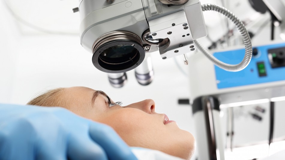
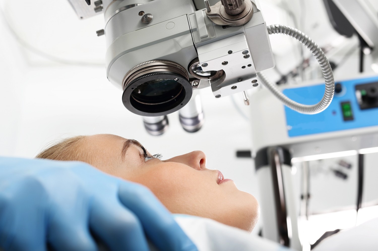
Researchers at the International Centre for Translational Eye Research (ICTER) have developed a prototype imaging system that can simultaneously track the cornea’s mechanical response at nine locations. The technology marks an important step toward more accurate, non-contact assessment of corneal biomechanics and could improve early detection and monitoring of disorders such as keratoconus.
The cornea is the transparent front surface of the eye and the first structure that bends incoming light. Its shape and optical properties are essential for sharp vision. Yet the cornea is much more than a transparent window. It is a complex, layered tissue whose mechanical properties influence how it responds to pressure, external forces, and even subtle changes in its own structure.
Understanding these biomechanical properties has become increasingly important in modern ophthalmology. They can reveal changes that appear before visible structural abnormalities develop and help clinicians assess disease progression or treatment outcomes. However, accurately measuring corneal biomechanics remains technically challenging.
Researchers from ICTER, operating at the Institute of Physical Chemistry of the Polish Academy of Sciences, have now developed a new optical coherence tomography (OCT) system designed specifically for this purpose. Their study, Single-shot, depth-encoded multiplexed OCT for multi-spot tracking of induced transient corneal dynamics, published in Biomedical Optics Express, describes a prototype capable of recording how the cornea deforms after a short air pulse – all within a single measurement.
The ICTER authors of the publication, both current and former members of the Centre, are Karol Karnowski, Jadwiga Milkiewicz, Onur Cetinkaya, Angela Pachacz, Andrea Curatolo, Kamil Liżewski, Dawid Borycki, and Maciej Wojtkowski. The project was carried out in collaboration with researchers from Nicolaus Copernicus University in Toruń, the Collegium Medicum of Nicolaus Copernicus University in Bydgoszcz, the University of Liverpool, the Instituto de Óptica Daza de Valdés in Madrid, and the University of Rochester.
Looking beyond conventional corneal measurements
Modern ophthalmic instruments routinely measure corneal thickness, curvature, and overall shape. Many also assess how the cornea responds to a gentle mechanical stimulus. One of the best-known examples is the non-contact air-puff test, widely used to measure intraocular pressure. During the examination, a brief pulse of air causes a slight deformation of the cornea, which is then analysed by the instrument.
While this approach has become a clinical standard, it has an important limitation. Mechanical weakening of the cornea is not always uniform.
In keratoconus – a progressive disease that causes the cornea to thin and bulge outward – localised areas of tissue may become mechanically weaker long before conventional imaging reveals obvious structural changes. As the disease progresses, patients may experience worsening vision, increasing astigmatism, greater sensitivity to light, and growing difficulty achieving good visual correction with glasses or contact lenses. Advanced cases may ultimately require specialised treatment or even corneal transplantation.
Existing biomechanical assessment methods typically evaluate the overall corneal response or analyse deformation within a single cross-sectional plane. Although these approaches provide valuable clinical information, they may overlook subtle, localised abnormalities occurring away from the central measurement axis.
The ICTER team set out to overcome this limitation.
Instead of analysing the cornea one location at a time, their system measures its response simultaneously at nine positions – one central point and eight surrounding peripheral locations. This makes it possible not only to determine how much the cornea deforms, but also to identify precisely where the tissue responds most strongly and whether that pattern may indicate local biomechanical weakening.
“The cornea is one of the eye’s primary optical elements responsible for focusing light onto the retina, so not only its shape but also its mechanical properties are crucial. It is believed that the mechanical state of the cornea changes in certain conditions, such as keratoconus, before any alterations in its shape become apparent. By simultaneously measuring the cornea’s response to an air-puff stimulus, our system can assess this mechanical asymmetry, offering the potential to detect pathological changes earlier than methods based solely on corneal geometry,” says Dr Karol Karnowski from ICTER.
Capturing nine measurement points in a single shot
The new technology builds on optical coherence tomography (OCT), one of the most widely used imaging techniques in ophthalmology. OCT provides high-resolution cross-sectional images of the eye using light rather than sound, making it the optical counterpart of ultrasound. It has become indispensable in retinal imaging and anterior segment diagnostics because it can visualise ocular tissues quickly, non-invasively, and with micrometre-scale resolution.
In this project, however, the ICTER researchers pushed the technology beyond conventional OCT imaging.
Rather than illuminating the cornea with a single probing beam, they designed an optical system in which several beams strike the tissue simultaneously. Each beam corresponds to a different measurement location. Instead of acquiring these locations sequentially, the signals are separated using depth encoding, allowing information from multiple points on the cornea to be recorded within a single OCT acquisition.
The advantage is straightforward but significant. Conventional scanning systems collect data point by point, meaning that measurements from different locations are acquired at slightly different moments in time. For relatively static tissues, this is rarely a problem. Corneal deformation following an air puff, however, is a rapid, transient event lasting only a few tens of milliseconds. Even tiny eye movements, blinking, fluctuations in the tear film, or the inherent delay of sequential scanning can introduce measurement errors.
By recording all nine locations simultaneously, the ICTER system eliminates these temporal inconsistencies. Every point is measured during the same mechanical event, providing a much more faithful representation of how the cornea responds to external force.
The prototype operates at exceptionally high speed. It records corneal motion every 10 μs, corresponding to 100,000 measurements per second across all nine locations simultaneously.
The researchers also investigated what would happen if the temporal resolution were reduced. When sampling was degraded to 250 μs, the error in estimating the magnitude of the asymmetry vector reached approximately 20%. At 1 ms, the error approached 70%. The effect was even more pronounced when estimating the direction of the asymmetry vector: the angular error increased to 17.5° at 250 μs and as much as 47.5° at 1 ms.
These findings demonstrate that temporal resolution is not merely a technical specification – it fundamentally determines measurement accuracy. Slower acquisition rates may not only underestimate or overestimate the extent of biomechanical abnormalities but also misidentify the region where the cornea is weakest.
“The corneal deformation we aim to capture to assess its mechanical asymmetry lasts only about 20 milliseconds, and the measurement cannot be repeated. This makes the simultaneous acquisition of the corneal response at multiple locations the key feature of our method. Achieving this requires very high temporal resolution – much like a camera capable of capturing an extremely high number of frames per second. Existing techniques have either lacked sufficient temporal resolution or have been limited in their spatial coverage,” explains Dr Karol Karnowski.
For this reason, simultaneous acquisition represents one of the key innovations of the new system. Instead of reconstructing corneal behaviour from measurements taken fractions of a millisecond apart, the technology captures the entire biomechanical event as it unfolds.
The result is a more reliable representation of corneal mechanics and a significantly improved ability to detect subtle local biomechanical differences that may otherwise remain hidden.
The asymmetry vector: turning complex motion into a clinically meaningful biomarker
Recording corneal deformation at nine locations simultaneously generates a wealth of biomechanical information. To make these data clinically useful, the researchers developed a parameter called the asymmetry vector.
The concept is straightforward. The system compares corneal deformation at eight peripheral locations with the response at the centre and between opposing measurement points. If one region deforms more than another, this indicates biomechanical imbalance within the tissue.
The asymmetry vector describes this imbalance using two parameters. Its magnitude reflects how large the biomechanical difference is, while its direction indicates where the greatest deformation occurs. Together, they provide a concise representation of corneal behaviour that is easy to compare between patients and across follow-up examinations.
One way to think about the asymmetry vector is as a biomechanical compass. Rather than simply indicating that the cornea behaves asymmetrically, it points toward the region where that asymmetry is most pronounced.
This could prove particularly valuable in keratoconus.
In keratoconus, tissue weakening, thinning, and protrusion rarely occur exactly in the centre of the cornea. Instead, the disease typically develops locally, affecting only part of the tissue in its earliest stages. As a result, the average biomechanical response of the entire cornea may appear relatively normal even though one region has already become mechanically compromised.
The researchers demonstrated that the asymmetry vector consistently pointed toward the area affected by keratoconus. In these patients, the vector direction corresponded to the region where the cornea was thinner, steeper, and showed abnormal posterior elevation.
The authors note that the vector did not perfectly overlap with maps generated by commercially available corneal tomography systems. This was expected, since the two techniques measure different aspects of the tissue.
Conventional topography and tomography describe corneal shape and anatomy. The new OCT-based approach measures how the tissue behaves mechanically when subjected to a brief air pulse. Rather than competing with existing diagnostic methods, the researchers see these measurements as complementary.
“The asymmetry vector provides a way to translate the cornea’s rapid and complex motion into a single parameter that can be compared across patients and between successive examinations. From a clinical perspective, the key question is whether it can identify the predominant direction of biomechanical weakening. In the future, such a parameter could support risk assessment and disease monitoring,” explains Jadwiga Milkiewicz, MSc, from ICTER.
One air pulse, two distinct corneal deformations
While validating the prototype, the researchers observed an unexpected phenomenon. Instead of deforming only once after a single air pulse, the cornea occasionally exhibited two distinct deformation events. The team named this previously unreported behaviour the dual-indentation response. The discovery emerged after the researchers modified the air-delivery system.
To allow measurements not only at the corneal apex but also across the peripheral measurement points, they increased the diameter of the air nozzle from 1.5 mm to 3.7 mm. The broader nozzle produced a wider air pulse while reducing its peak force by approximately half. Importantly, the pulse duration remained unchanged at 8 ms. Although the stimulus became weaker, the biomechanical response became more complex.
Instead of a single indentation followed by recovery, the cornea developed two clearly distinguishable deformation phases after the same air pulse. This suggests that widening the air stream changes how mechanical force is distributed across the corneal surface, revealing dynamic behaviour that conventional measurements may fail to capture.
Because all nine locations were recorded simultaneously and at extremely high temporal resolution, the researchers were able to observe this phenomenon directly rather than infer it retrospectively from sequential scans.
“Before the data can be analysed, they must be carefully prepared – cleaned, standardised, and corrected for measurement artefacts. The reliability of the conclusions depends heavily on the quality of this preprocessing stage, which is why we devote as much attention to it as to the measurement itself,” says Dr Dawid Borycki.
Although the biological mechanisms underlying this response remain to be investigated, the observation illustrates the value of high-speed, simultaneous biomechanical imaging. By capturing the entire event rather than reconstructing it from sequential measurements, the system can reveal subtle dynamic phenomena that might otherwise remain hidden.
What could this mean for patients?
Although the current study is primarily a technical proof of concept, the technology has clear clinical potential.
Because the system combines optical coherence tomography with a brief air pulse – both already familiar in ophthalmic practice – it could be integrated into future diagnostic devices without fundamentally changing the patient experience. The innovation lies not in the examination itself, but in how the corneal response is recorded and analysed.
One of the most promising applications is the earlier detection of keratoconus. In many patients, biomechanical changes precede visible structural abnormalities. A system capable of identifying subtle, localised weakening of the cornea could therefore detect disease at an earlier stage than methods that rely solely on morphology.
The technology could also support long-term monitoring of disease progression and provide an objective way to assess treatment outcomes. This may be particularly valuable following corneal cross-linking, a procedure designed to strengthen corneal tissue and slow the progression of keratoconus.
Another potential application is refractive surgery planning. Procedures such as LASIK and other forms of laser vision correction require careful assessment of corneal biomechanics to minimise the risk of postoperative complications. More detailed biomechanical information could help clinicians identify patients who are not suitable candidates or refine treatment planning for those who are.
The authors emphasise, however, that the present study focused on demonstrating the capabilities of the technology rather than validating its clinical performance.
A separate clinical dataset has already been collected, including healthy volunteers as well as patients with early and mild forms of keratoconus. The results of this validation study will be presented in a future publication.
Like any emerging technology, the current prototype also has limitations.
At present, analysis requires manual segmentation of the anterior corneal surface in OCT images. While this approach is robust and appropriate for research, it is too time-consuming for routine clinical use. The ICTER team is therefore developing automated analysis pipelines, including machine learning-based algorithms that could substantially accelerate image processing.
Accurate eye positioning remains another technical challenge. Small eye movements, incomplete alignment, or blinking can reduce signal quality and affect measurement accuracy. Future versions of the system may therefore incorporate pupil monitoring, real-time alignment feedback, and eye-tracking technology to improve robustness during clinical examinations.
Beyond the cornea
Although the study focuses on corneal biomechanics, the researchers believe that the underlying measurement principle could have applications far beyond ophthalmology.
The work demonstrates that simultaneous multi-point OCT imaging can capture extremely rapid, non-repeatable mechanical events in light-scattering biological tissues – situations in which conventional sequential scanning may introduce significant temporal errors.
This concept could ultimately prove valuable for studying other soft tissues where fast mechanical processes are difficult to capture reliably with existing imaging techniques.
The prototype described in this study represents an important step toward that broader vision. By combining ultrafast acquisition with simultaneous multi-point measurements, the ICTER team has shown that OCT can provide not only detailed structural images but also new insights into tissue biomechanics.
As the technology continues to mature and undergo clinical validation, it could become a valuable addition to the ophthalmic diagnostic toolbox, enabling clinicians to detect biomechanical abnormalities earlier, monitor disease progression more precisely and make better-informed treatment decisions.
------------------------------------------------------------------------------------------------------
Karol Karnowski, Jadwiga Milkiewicz, Onur Cetinkaya, Angela Pachacz, Ewa Mączyńska-Walkowiak, Patryk Młyniuk, Andrea Curatolo, Kamil Liżewski, Ahmed Abass, Dawid Borycki, Bartłomiej Kałużny, Susana Marcos, Ahmed Elsheikh, Ireneusz Grulkowski, and Maciej Wojtkowski (2026). Single-shot, depth-encoded multiplexed OCT for multi-spot tracking of induced transient corneal dynamics. Biomedical Optics Express.
- Author: Scientific Editor Marcin Powęska
- Photo source: Fot. Depositphotos
- Date: 27.07.2026